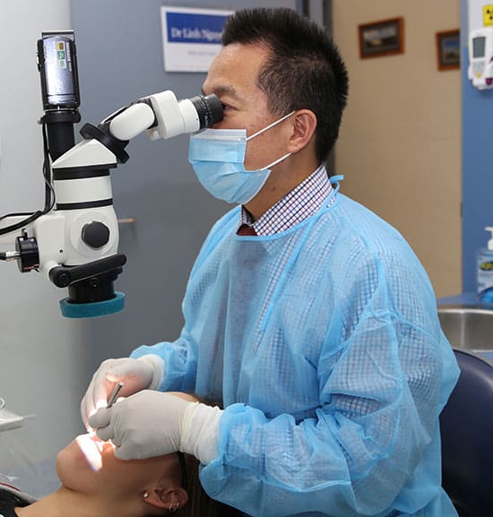
Dental Microscope
The dental microscope is used to promote consistency and excellence in dental procedures. The microscope magnifies and illuminates patient’s mouths, allowing for greater ease and precision for the dentist in examining a patient and performing procedures. Some dental procedures occur in a very small area: for example, the root canal procedure occurs in a small area of the mouth and the use of a microscope not only enables the dentist to view the area with more ease, but also displays the area in enough detail to show the dentist issues that would not be visible to the naked eye, such as a tooth that has more roots and/or canals than expected. The dental microscope is also a great help for locating broken roots in many surgical removals of broken roots in the dental sockets. This increases the chance of a successful and thorough procedure.
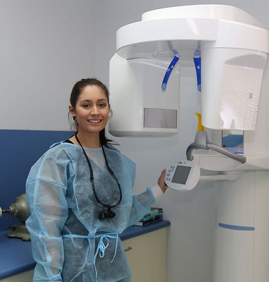
Cone Beam and OPG X-ray Machine
A Cone Beam is a relatively new type of dental imaging. Cone Beams produce 3D images in high resolution scans, which also have the benefit of lower radiation exposure for patients. Cone Beam imaging is done by placing a patient in the scanner, while the machine rotates once all the way around a patient’s head. The process takes 10-15 seconds on average and the data is then processed into a 3D image. The benefit of a Cone Beam image is in the detail, which allows the dentist to view the image from any angle to make an accurate diagnosis. The OPG X-ray Machine, like Cone Beam imaging, improves diagnostics by providing the dentist with more visibility and detail of a patient’s dental issues. The patient stands with their face resting on a small shelf and bites gently on a sterile mouthpiece – this ensures the patient’s head is steady for the x-ray. The OPG X-ray focuses on the lower face, teeth, jaw joints and maxillary sinuses, which can allow dentists to see the position and growth of teeth that have not even emerged yet. The OPG can be used to plan orthodontic treatment, to assess wisdom teeth and for a general overview of the teeth and surrounding bones.
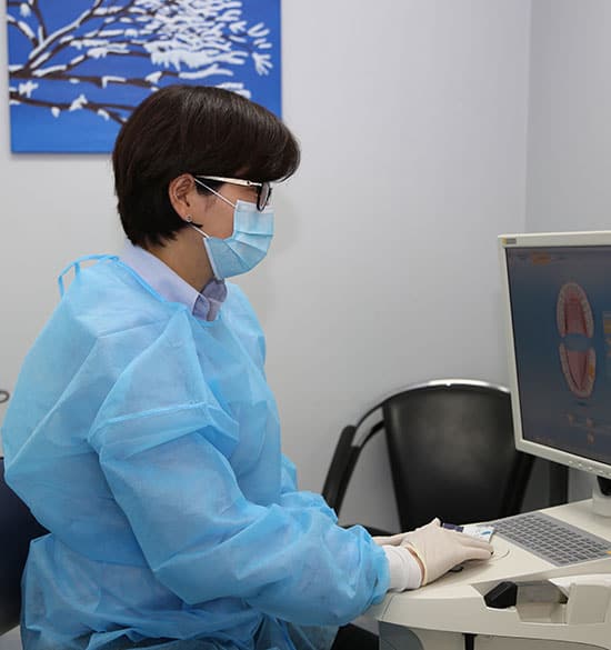
CEREC Machine
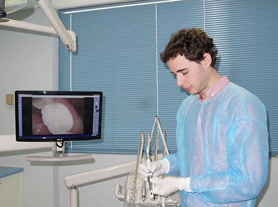
Intra-Oral Camera
An intra-oral camera is a small device that looks like a pen, connected to a dentist’s computer and large screen television to allow the dentist to see the inside of the mouth in greater detail. The light connected to the camera allows the dentist to see cavities and/or fractures, which would otherwise be invisible to the naked eye, allowing for greater accuracy and detail in diagnosing any issues. In addition, the intra-oral camera also has the benefit of allowing a patient to see the inside of their own mouth, through the connection to the computer and TV. The patient is able to see issues described by the dentist and as well as the overall state of their mouth.